Urinary bladder
| BLADDER | |
|---|---|
 |
|
| 1. Human urinary system: 2. Kidney, 3. Renal pelvis, 4. Ureter, 5. Urinary bladder, 6. Urethra. (Left side with frontal section) 7. Adrenal gland |
|
 |
|
| Male Bladder Makeup | |
| Latin | vesica urinaria |
| Gray's | subject #28 1227 |
| Artery | Superior vesical artery Inferior vesical artery Umbilical artery Vaginal artery |
| Vein | Vesical venous plexus |
| Nerve | Vesical nervous plexus |
| Lymph | external iliac lymph nodes, internal iliac lymph nodes |
| Precursor | urogenital sinus |
| MeSH | Bladder |
| Dorlands/Elsevier | Urinary bladder |
In anatomy, the urinary bladder is the organ that collects urine excreted by the kidneys prior to disposal by urination. A hollow[1] muscular, and distensible (or elastic) organ, the bladder sits on the pelvic floor. Urine enters the bladder via the ureters and exits via the urethra.
Embryologically, the bladder is derived from the urogenital sinus and, it is initially continuous with the allantois. In males, the base of the bladder lies between the rectum and the pubic symphysis. It is superior to the prostate, and separated from the rectum by the rectovesical excavation. In females, the bladder sits inferior to the uterus and anterior to the vagina. It is separated from the uterus by the vesicouterine excavation. In infants and young children, the urinary bladder is in the abdomen even when empty.[2]
Contents |
Detrusor muscle
The detrusor muscle is a layer of the urinary bladder wall made of smooth muscle fibers arranged in spiral, longitudinal, and circular bundles. When the bladder is stretched, this signals the parasympathetic nervous system to contract the detrusor muscle. This encourages the bladder to expel urine through the urethra.
For the urine to exit the bladder, both the autonomically controlled internal sphincter and the voluntarily controlled external sphincter must be opened. Problems with these muscles can lead to incontinence. If the amount of urine reaches 100% of the urinary bladder's capacity, the voluntary sphincter becomes involuntary and the urine will be ejected instantly.
The urinary bladder usually holds 100000-200000 mL of urine; a full adult bladder holds about 500mL of urine, 15 times its empty volume. Not all specialists accept these values, some say a urinary bladder can hold ca. 1000 mL, but it is different from person to person. As urine accumulates, the rugae flatten and the wall of the bladder thins as it stretches, allowing the bladder to store larger amounts of urine without a significant rise in internal pressure.[3]
The desire to urinate usually starts when the bladder reaches around 125% of its working volume. At this stage it is easy for the subject, if desired, to resist the urge to urinate. As the bladder continues to fill, the desire to urinate becomes stronger and harder to ignore. Eventually, the bladder will fill to the point where the urge to urinate becomes overwhelming, and the subject will no longer be able to ignore it.
Since the urinary bladder has a transitional epithelium, it does not produce mucus.[4]
Fundus
The fundus of the urinary bladder is the base of the bladder, formed by the posterior wall. It is lymphatically drained by the external iliac lymph nodes. The peritoneum lies superior to the fundus.
Disorders
Disorders of or related to the bladder include:
- Bladder cancer
- Bladder exstrophy
- Bladder infection
- Bladder spasm
- Bladder sphincter dyssynergia, a condition in which the sufferer cannot coordinate relaxation of the urethra sphincter with the contraction of the bladder muscles
- Bladder stones
- Cystitis
- Hematuria, or presence of blood in the urine, is a reason to seek medical attention without delay, as it is a symptom of bladder cancer as well as bladder and kidney stones.
- Interstitial Cystitis
- Overactive bladder, a condition which affects a large number of people.
- Urinary incontinence
- Urinary retention
In other animals
The bladder of fishes is generally small, and is not homologous with that of tetrapods. In lobe-finned fish, primitive ray-finned fish, and in many female cartilaginous fish, the bladder is formed from the fused ends of the archinephric ducts, counterparts of the mammalian ureters. In male sharks and rays, however, these ducts are only used for passage of sperm, and do not form a bladder. However, a bladder is sometimes found in these fish, developed from additional urinary ducts posterior to the archinephric ducts.[5]
In teleosts and lampreys, the bladder forms from part of the wall of the cloaca, where it is present at all, although the archinephric ducts are usually also involved in its formation. Like that of other fish, it is not lined by the transitional epithelium found in tetrapods.[5]
The bladder of amphibians and reptiles is a pocket in the cloaca, and is usually not connected with the urinary ducts at all. Only in mammals, most of which have no cloaca, does the bladder take on the general form seen in humans. Many reptiles, including snakes and crocodilians, have no bladder, and the only bird species to have a bladder is the ostrich.[5]
Uses
Besides its normal use to the possessor, animal bladders (usually pig bladders) have been used to make balls (such as footballs) and even a musical instrument, the bumbass.
See also
- Artificial bladder
- Bladder augmentation
- Neurogenic bladder
- Ureterocele
- Urodynamics
- Uvula of urinary bladder
- Vesicouretic reflux
References
- ↑ Howard A. Werman, MD and Keith J. Karren, PhD.
- ↑ Moore, Keith L.; Dalley, Arthur F, Clinically Oriented Anatomy, 5th Edition, 2006 Lippincott Williams & Wilkins
- ↑ Marieb, Mallatt Human Anatomy 5th Edition Pearson International Edition Chapter 23 p700
- ↑ T . Chin , C . Liu , H . Tsai , C . Wei. Vaginal reconstruction using urinary bladder flap in a patient with cloacal malformation
- ↑ 5.0 5.1 5.2 Romer, Alfred Sherwood; Parsons, Thomas S. (1977). The Vertebrate Body. Philadelphia, PA: Holt-Saunders International. pp. 378–379. ISBN 0-03-910284-X.
External links
- Histology at KUMC epithel-epith09 "Urinary Bladder"
- Organology at UC Davis Urinary/mammal/bladder/bladder1 - "Mammal, bladder (LM, Medium)"
- Virtual Slidebox at Univ. Iowa Slide 445
- SUNY Labs 43:07-0100 - "The Female Pelvis: The Urinary bladder"
- SUNY Labs 44:04-0103 - "The Male Pelvis: The Urinary bladder"
Additional images
 Structure of the penis |
 Organs of the female reproductive system. |
 Coronal section of pelvis, showing arrangement of fasciæ. Viewed from behind. |
 Dissection of side wall of pelvis showing sacral and pudendal plexuses. (Bladder visible at lower left.) | |
 The peritoneum of the male pelvis. |
 Median sagitta section of male pelvis. |
 Male pelvic organs seen from right side. |
 Median sagittal section of female pelvis. |
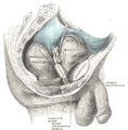
 The interior of bladder. |
 Vertical section of bladder wall. |
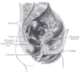
 Fundus of the bladder with the vesiculæ seminales. |
 Vertical section of bladder, penis, and urethra. |
 Female pelvis and its contents, seen from above and in front. |
 Topography of thoracic and abdominal viscera. |
 The bladder can be seen highlighted in yellow in the illustration. |
 Layers of the urinary bladder wall and cross section of the detrusor muscle. |
|
|||||||||||||||||||||||||||||||||||||||||||||||||||||||||||||
|
||||||||||||||||||||||||||||||||||||||||||||